Scoliosis

You Are Not Alone
Scoliosis Treatment
Scoliosis Treatment
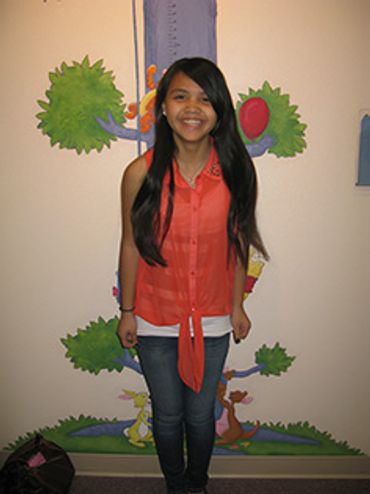
Got Scoliosis? "Curvy Girls® support group has your back.

Scoliosis Treatment
Scoliosis Treatment
Scoliosis Treatment
There are currently no medications to treat scoliosis, but there are two other ways.

The Rigo Cheneau
Scoliosis Treatment
Our New Technology System
This is our brace of choice because over 30 years it has proven & out performed others.

Our New Technology System
Our New Technology System
Our New Technology System
We use a Scoliosis scanning machine, CAD Software System & The CANFIT-PLUS CARVER.

We Provide Custom Care
Our New Technology System
We Provide Custom Care
We treat patients with scoliosis by creating custom braces with new technology.

Scoliosis Treatment
We Can Help
There are currently no medications to treat scoliosis, nor can its onset be prevented. When scoliosis is detected, the doctor may refer the patient to an orthopedic spinal specialist for evaluation and treatment. This may consist of periodic examinations, including standing X-rays as needed to determine if the curve is increasing in size. If scoliosis is identified early, large curves may often be prevented by wearing a brace. Severe curves may require surgical treatment. There are two ways to treat scoliosis:
Custom bracing: If the curvature is below 45 degrees, a surgical operation is not necessary to treat scoliosis.
Surgical: If the curvature is above 45 degrees, a surgical operation is currently the only way to treat scoliosis.
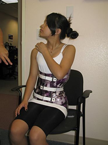
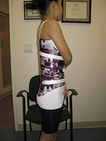
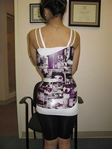
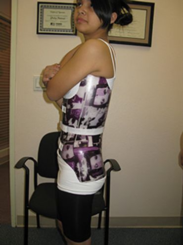
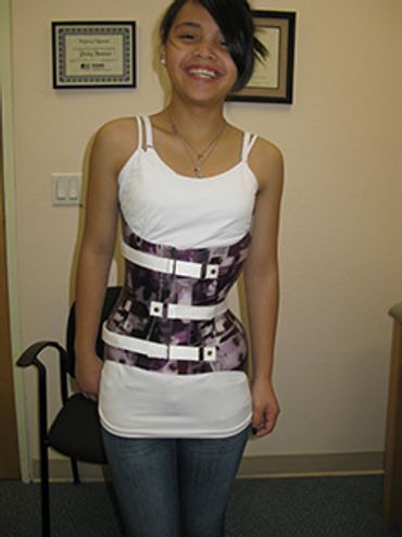
The Rigo Cheneau Brace Program
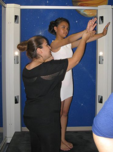
Step 1: Initial evaluation/scanning
Step 1: Initial evaluation/scanning
Step 1: Initial evaluation/scanning

At this appointment, X-Ray and report from the Orthopedic Physician are reviewed. After we review the x-rays we talk a scanning and measurements. Then we get to work on preparing a custom made brace. We also address concerns of our patients and provide information on scoliosis.
The Rigo Cheneau Brace
Step 1: Initial evaluation/scanning
Step 1: Initial evaluation/scanning
The Rigo Cheneau Brace is our brace of choice because over 30 years of experience have proven to us that it out performs other braces.
Carving
Step 1: Initial evaluation/scanning
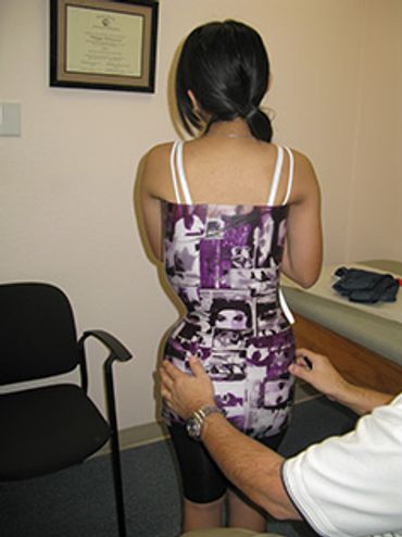
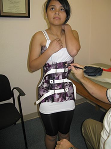
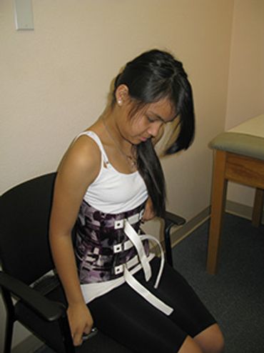
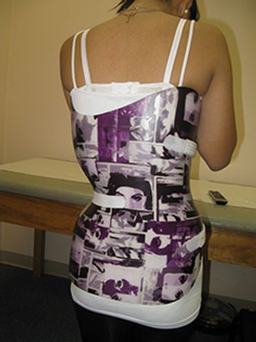
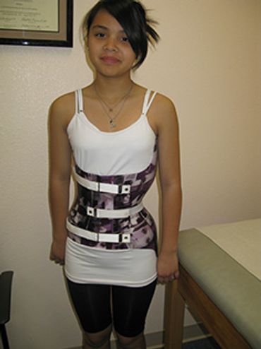
Step 2: Fitting of the brace
....
Step 2: Fitting of the brace
Step3: First x-ray in the brace
Step 2: Fitting of the brace
This is a long appointment (typically 2 hours) because we want to make sure each brace feels and fits right. The first time our patients wear the brace, it may look and feel weird, but we aim to make it as comfortable as possible. We offer support suggestions and clothing advice.
Step3: First x-ray in the brace
Step3: First x-ray in the brace
Step3: First x-ray in the brace
(Timing: 2-3 weeks after step 2) The brace should have been worn full time if possible. The x-ray in brace and the report from the Orthopedic physician is reviewed and base on that adjustments are made to the brace.
Step4: Follow up appointment
Step3: First x-ray in the brace
Step3: First x-ray in the brace
(Timing: every 6-8 weeks) Patients growth, brace fit and use must be monitored.
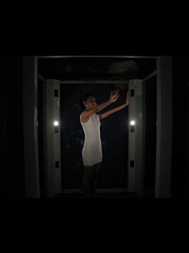
Our New Technology Scoliosis Scanning System
3D Measurement Device
The CANFIT-PLUS CARVER
3D Measurement Device
he 3D scanning device is quick and accurate. Optical casting is non-invasive, eliminates plaster casts, and the need for prolonged direct contact with the patient. Called ComfORTAC, it acquires 360 degrees measurements of a patients body and legs using structured light projection. The result scan is used by our CAD software to design orthopedic devices. Exact physical reference points are recorded directly from their position on the patient. These measurements are exactly reproduced, and stored to the computer.
- The 3D measuring device takes two pictures from the patient: frontal and profile. The software automatically finds the silhouette and calculate the 3D model.
- By acting directly on the patient’s contour, the 3D model is automatically modified. The resulting model fits the patient’s contour perfectly
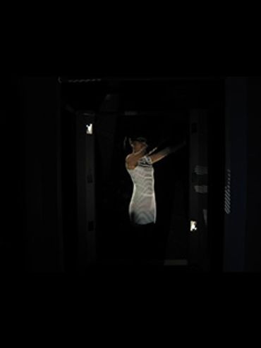
CAD Software System
The CANFIT-PLUS CARVER
3D Measurement Device
This system provides an integrated suite of tools for acquiring shape data, designing and modifying shapes, and carving positive models.
- Accurate and efficient – CAD software models data from the 3D scan and enables the operator to make appropriate corrections.
- Working with an actual 3D image of the patient, measurements are reproduced exactly to create a viewable virtual cast.
- The electronic data is automatically stored. 3D monitoring and adjustments are made if needed as the treatment progresses.
The CANFIT-PLUS CARVER
The CANFIT-PLUS CARVER
The CANFIT-PLUS CARVER
The CANFIT-PLUS CARVER is designed to carve industry standard polyurethane foam blanks, handling shapes and sizes ranging from small upper extremities to very large spinal shapes.
- The 3D model from the CAD system is sent to another machine, the CANFIT-PLUS milling machine, a computer numerically controlled (CNC) carver which produces a polyurethane positive model of the socket or brace.
- The unique software enables environmentally safe and economical reuse of polyurethane blocks.
- The carver features advanced design, large capacity and extreme accuracy.

We Provide Custom Care
Custom Braces
At O&P In Motion Inc., we treat patients with scoliosis using custom braces. Most facilities take a plaster mold to cast the brace. We use a Scoliosis scanning machine instead! The advantages in using the Scoliosis scanning system are:
- Faster than plaster casting
- More precise and accurate
- No plaster mold impression/comfort for the patient
- The measurement is exactly reproduced
- The professional works on the actual 3D image of the patient (not model)
The Process